|
For Patients
Barrett's Oesophagus Detection and Diagnosis
Upper Endoscopy
Most commonly, Barrett's Oesophagus is diagnosed during an upper endoscopy procedure, or also known as esophagogastroduodenoscopy (EGD).
The endoscopy procedure consists of a thin, flexible tube that is guided down your throat.
The tube, known as an endoscope, has a video lens and light at its tip that transmit images to a video monitor nearby. This allows the doctor to visually inspect and capture images of the tissue of your oesophagus. The endoscopy procedure is typically performed by a gastroenterologist or a gastrointestinal surgeon in an outpatient office, ambulatory surgery center or hospital endoscopy suite. The procedure typically takes less than 30 minutes to perform and the patient goes home following the procedure.
There are new thin endoscopes that allow the physician to pass an endoscope through the patient's nose to quickly and conveniently check the patient for Barrett's oesophagus.
There are also new small capsules with built-in cameras that the patient may swallow and have a physician screen them for Barrett's oesophagus.
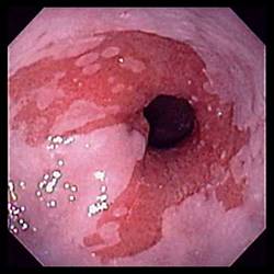
Barrett's oesophagus appears as red-colored tissue, as compared to the normal pink colored oesophagus lining.
 |
| Example of the image seen from the endoscope. The darker red oesophageal tissue consists of Barrett's cells. |
Biopsy
Once diagnosed with Barrett's oesophagus visually, your doctor will also obtain a tissue sample, or biopsy, from your oesophagus during an upper endoscopic procedure. The tissue from the biopsy is analyzed under a microscope to confirm the diagnosis and determine the stage of the Barrett's oesophagus.
|
Barrett's Disease
|
Barrett's Oesophagus
|
Barrett's Esophagus
|
Barrett's 0esophagus
|
Barrett's Esophagitis
|
|
Barratts Disease
|
Barratts Oesophagus
|
Barretts Esophagus
|
Barretts 0esophagus
|
Oesophageal Cancer
|
|
|
|

